
Lesson 3 - Diagnosing Celiac Disease
3.3 Special situations
 Testing for the CeD risk variants HLA-DQ2/DQ8 is not necessary in a normal diagnostic work-up, regardless the CeD diagnosis is done with biopsies or without, based on serology only. The specificity is very low. However, HLA-typing may be helpful to exclude the risk for or the diagnosis of CeD due to its negative predictive value of almost 100% (Unit 1, lesson 1.3).
Possible situations in which genetic testing may be helpful (Husby et al. 2012; Husby et al. 2019; Rubio-Tapia et al. 2013):
- Children with family member with proven CeD: In about 20% of first-degree relatives of CeD patients, HLA-DQ2/D8 is negative and consequently, no further follow-ups for CeD serology is necessary. If they are positive, regular follow-up of TGA-IgA (or IgG antibodies in the case of IgA deficiency) are recommended! In newborns blood sampling for HLA-testing may be taken from the umbilical cord.
- Young children with Down syndrome (Trisomy 21) or Turner Syndrome: the prevalence of HLA-DQ2/DQ8 is similar as in the general population, this means a 60 to 70% are negative and do not need to be further screened for CeD auto-antibodies. If positive, regular follow-up of TGA-IgA (or IgG antibodies in the case of IgA deficiency) is recommended!
- People who have started a gluten-free diet without prior testing for auto-antibodies or without (properly performed) upper endoscopy: Since both auto-antibodies and the mucosa usually normalize within weeks to months after starting a gluten free diet, a reliable diagnosis cannot be made using serology tests or intestinal biopsy. HLA testing allows the exclusion of CeD in individuals that are negative for HLA-DQ2 and -DQ8. If the test is positive for the HLA risk markers, only a gluten challenge with further testing for auto-antibodies and in most cases also biopsies will allow us to confirm or exclude CeD (CR Unit 2, lesson 1).
- Patients with mucosal damage confirmed by biopsies but with negative auto-antibodies: If the HLA-DQ2 and –DQ8 risk markers are negative, CeD is extremely unlikely and another disease must be considered as responsible for mucosal inflammation and damage. If the test is positive, the situation remains unclear. Repeated biopsies on a gluten-containing diet (respectively after gluten challenge) are needed to confirm or exclude celiac disease.
Unlike blood tests and endoscopy, the genetic test for celiac disease can be performed regardless of the patient’s gluten consumption. Depending on the country and the laboratory, the expensive HLA-testing may however not always be covered by the health insurance.
Diagnosing "celiac disease of the skin" - Dermatitis herpetiformis Duhring (DH)
The gold standard for diagnosis of DH is the detection of granular IgA deposits by the method of direct immunofluorescence microscopy in the skin of patients (Rose et al. 2009, Görög et al, 2021). The biopsy should be taken from clinically normal-appearing skin near the lesion (within 1 cm or less). The reason is that lesional skin may be devoid of IgA and can lead to a false negative result. Direct immunofluorescence microscopy can only be performed from a frozen tissue sample without fixation in formalin or formaldehyde. CeD-specific serology (TGA or EMA) should also be analysed.
If DH is diagnosed in the skin biopsy and the serology is confirming CeD, the patient may not need an intestinal biopsy (Bolotin & Petronic-Rosic 2011, Görög et al, 2021).
In cases where DH is suspected, the patient should be referred to a dermatology center with experience which is experienced in this condition.

Gluten challenge
Nowadays a gluten challenge is only rarely performed. However, there are situations in which it is obligatory.
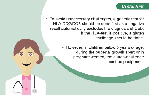
If a child or adult is already on a gluten-free diet without a proper diagnosis of CeD, or if there is doubt about the initial diagnosis, a gluten challenge with or without prior testing for HLA risk markers is recommended. However, in children under the age of 5 years, during the pubertal growth spurt, during pregnancy or when a pregnancy is anticipated, a gluten challenge should be postponed (Husby et al. 2012; Celiac Disease Foundation 2017).
During the challenge, some, but not all, patients may develop symptoms. However, all patients with CeD will produce auto-antibodies and usually villous atrophy. The duration of the gluten challenge may vary from patient to patient: some will react with symptoms within days followed by increased titres of TGA antibodies within a few weeks. In others it may take several months or even years until the disease re-occurs.
How is a gluten challenge performed?
During a gluten challenge, the daily intake of gluten containing food should be of normal amount, at least equivalent of 3g gluten in children (e.g. ~1-2 slices of regular bread or one plate of pasta or pizza) (Husby et al. 2012) (Rubio-Tapia et al. 2013), and up to 15g in adolescents and adults (Felber et al. 2014). Alternatively, gluten powder may be mixed in regular food to help avoid familiarization with the taste of gluten containing products. This may be particularly important in children who may have problems returning to a gluten-free diet after a gluten challenge and confirmation of the diagnosis.
Blood should be taken to test for auto-antibodies (TGA-IgA) when symptoms occur. In the absence of symptoms, auto-antibodies should be determined after 6 to 8 weeks, and if still negative every 3 to 6 months thereafter until 2 years after starting the challenge (Bruins JM 2013). Biopsies should be performed when TGA auto-antibodies become positive or symptoms occur. In the absence of auto-antibodies or symptoms, biopsies should be performed after 2 years on a gluten containing diet (Rubio-Tapia et al. 2013). Even if serology and biopsies after 2 years of gluten challenge do not indicate CeD, a later relapse cannot be completely excluded. Therefore these individuals should be tested periodically.