Lesson 3 - Diagnosing Celiac Disease
3.2 Biopsies and Histopathology
If the initial TGA-IgA test is positive, further investigations are always needed to confirm the diagnosis (Husby et al, 2020; NICE 2015). In many cases an upper endoscopy with biopsies is necessary to prove the mucosal lesions. In children and adolescents with very high TGA-IgA titres (higher than 10 times the upper limit of normal, ≥10 ULN) the diagnosis can be confirmed without biopsies if certain conditions are met.
If the first diagnostic step was an upper endoscopy with duodenal biopsies and histopathology shows clear villous atrophy, the diagnosis of CeD must be confirmed with a positive TGA-IgA (or IgG-based tests in case of IgA deficiency) before a gluten reduced/free diet is initiated. If CeD specific serology is negative, the diagnosis is unclear and further investigations including typing of HLA-DQ2/D8 as well as other differential diagnoses for villous atrophy must be considered (Unit 1, lesson 3.6)!
 Endoscopy
In adults, an upper endoscopy is usually performed by a gastroenterologist or nurse endoscopist, with or without local anaesthesia or under sedation.
In children and adolescents, endoscopy is usually done by a pediatric gastroenterologist or a surgeon. In younger children it requires general anesthesia and may therefore be more of a burden for the child and the family. Nonetheless it is also a safe procedure in children.
The endoscopist usually describes macroscopic abnormalities. However, only the histopathological examination of the duodenal biopsies is relevant for the diagnosis of CeD (Husby et al. 2020). Because villous lesions may not be equally distributed along the duodenum, the endoscopist should take at least 5 biopsies from different locations:
- at least 1 from the duodenal bulb
- at least 4 from the 2nd and 3rd part of the duodenum (Husby et al. 2020)
To ensure the correct orientation of the biopsies, it’s recommended to place the biopsies directly from the forceps on filter paper.
Please note: poor orientation of the specimen can lead to tangential cuttings and consequently artefacts, which the pathologist may not be able to assess or – even worse - may misinterpret them. This is one of the main pitfalls in the histopathological examination of CeD resulting in wrong diagnosis or incorrect exclusion of CeD (Taavela et al. 2013).
In case of contradicting results between serology and histopathology, the pathologist should be asked to review the biopsies again, considering that the correct orientation of the biopsies is important and re-orientation of wrongly oriented biopsy specimen is obligatory! In difficult cases, a reference pathologist should be involved and asked to pay special attention to the orientation and quality of the specimen.
Microscopic findings (histology)
The pathologist examines the duodenal biopsies for the following criteria (Celiac Disease Foundation 2017) (Dickson et al. 2006):
Orientation of the specimen
- Correct orientation allows direct examination of vertically cut villi and crypts
- Indirect respectively incorrect orientation does only allow indirect judgement or may even not allow any diagnostic statement.
Signs of Inflammation
- Increased number of intraepithelial lymphocytes (IEL)
Damage and changes of the mucosa, including:
- Shortening of the mucosal villi (villous atrophy)
- Lengthening of the crypts (crypt hyperplasia)
- Villous-crypt-ratio (a ratio of <2 in some parts of at least one biopsy indicates mucosal lesions)
Based on the findings, the pathologist should describe the degree of villous atrophy by using the Marsh or Marsh-Oberhuber staging (figure 12 and table 4) (Marsh 1992; Oberhuber et al. 1999). Marsh 0 (M0) indicates normal mucosa. Marsh 1 (M1) only means a non-specific increase of IEL. Marsh 2 (M2) is an intermediate stage defined by elongated crypts but normal length of the villi. Marsh 2 (M2) is nowadays considered to be indicative for CeD. Marsh 3 can be divided into three subtypes (M3A, 3B, 3C), ranging from shortened villi (M3A) to complete villous atrophy (M3C) with very deep crypts (Marsh-Oberhuber-staging).

Figure 12: The Marsh-(Oberhuber-) Classification of histologic findings.
From left to right, the graphic shows the variation from a normal morphology of the duodenal mucosa (Marsh 0) to an non-specific increase of IEL (Marsh 1), an elongation of the crypts (Marsh 2) and finally shortened to completely flat villi (Marsh 3A to C).
Source: FocusINCD own

Table 4: Histopathological features of the Marsh-Oberhuber staging with revised criteria for IEL count.
Important:
- A complete pathology report should always include a Marsh staging. Talk to your pathologist if the report is incomplete or discrepant to serology! Villous atrophy alone is not sufficient to diagnose CeD! In case of absence of CeD-specific autoantibodies despite a sufficient gluten-consumption of the patient, other differential diagnoses for villous atrophy need to be considered and further investigations are necessary!
- Some pathologists still use Marsh 4 to report too short villi, short crypts, hypoplastic changes. However, this is not associated with CeD but may be seen after irradiation, in nutrient deficiencies etc.
- Milder villous lesions do not mean that the person is more tolerant to gluten than patients with more severe atrophy (Husby et al. 2012). If the diagnosis of CeD is confirmed, a strict gluten exclusion diet is always the only therapy to achieve mucosal healing, regardless if the lesions were Marsh 2 or Marsh 3C.
Please note: The agreement between two pathologists regarding the diagnosis CeD yes or no is not perfect (discrepant results in 3 - 7%) (Villanacci et al, Werkstetter et al). Both, false positive and false negative reading may occur. Therefore, histopathology cannot serve as a reference standard and must be questioned in case of discrepant serology results.
The use of validated standard operating procedures (SOPs) with correct orientation and cutting of the duodenal specimen is of utmost importance for a correct interpretation of the mucosal changes.
Pitfalls in Histopathology
- The patient has already reduced amount of gluten and the mucosa has already recovered. In such cases the TGA-IgA antibodies are often negative as well. A gluten challenge is necessary in these cases.
- Villous atrophy can be patchy. If not enough biopsies were taken (at least 1 from duodenal bulb and 4 from 2nd or 3rd part of the duodenum) this may lead to false negative histology.
- Orientation of the biopsies: if the biopsies are not correctly oriented, they may not be suitable for reading (see Figure 13). Re-orientation and re-cuttings of the original biopsy blocks, in order to allow a correct interpretation, should be attempted.
- Low quality of the biopsies: if the biopsies are damaged (see figure 14 a and b), this may also cause misreadings and a new endoscopy with biopsies might be necessary before a gluten-free diet is started.

Figure 13: Impact of biopsy orientation.
Biopsy 1a and 2a (left side) both show apparently normal villi, but there are only cross-sections of the crypts visible, which indicates wrong orientation and cutting which does not allow any reading. However, pathologists have interpreted 1a and 2a as normal (Marsh 0 to 1). In the pictures 1b and 2b (right side) the same biopsies are shown which have been tilted and re-cut in correct orientation (perpendicular cutting). Now it is obvious that both 1b and 2b show villous atrophy and crypt hyperplasia and the same pathologists reported Marsh 3B to 3C for these biopsies. Both biopsies are clearly supporting a diagnosis of CD in combination with a positive serology, but misclassification can easily happen if the pathologist does not consider the correct orientation and cutting!
Source: FocusINCD with histology slides from Taavela et al. 2013
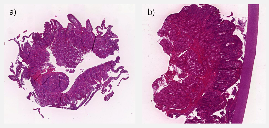
Figure 14: Examples of damaged biopsies which do not allow reading. Source: ProCeDE-study
Diagnosis without biopsies in children and adolescents
Very high TGA-IgA antibody titres (≥10 times upper limit of normal) have already a very high positive predictive value for CeD diagnosis (>99%). If the conditions are all fulfilled (see figure 15), the diagnosis of CeD in children and adolescents can be made with certainty without the need for histopathology (Husby et al 2012, 2020; Werkstetter et al 2017). This is also called the “no-biopsy approach”.
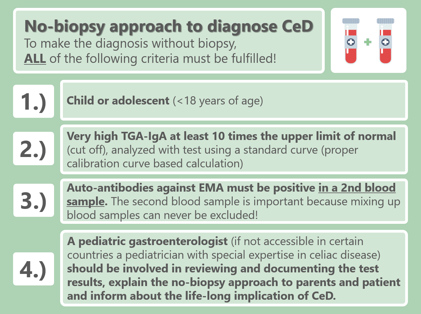
Figure 15: Diagnostic criteria for the diagnosis of CeD without biopsy.
Source: FocusINCD own with information from Husby et al. 2020
Please note: research is currently still investigating if the no-biopsy approach may also be applicable in adult patients. However, up to date (2021), the adult celiac guidelines have not yet included this possibility but revised guidelines may do so in the future.