
Lesson 2 - The Celiac Iceberg: Clinical Presentation & Symptoms
2.1 Different presentations of CeD
The presentation of CeD is variable and covers the whole spectrum of symptoms from severe malabsorption symptoms to other, less obvious signs or complete absence of any complaints (Riznik et al. 2021). Therefore, CeD is also called a clinical chameleon (Fasano et al. 2003).
Particularly those cases with less obvious clinical presentations often remain under the surface – only the tip of the iceberg is actually receiving a diagnosis (Unit 1 lesson 1.3 and figure 7).

Figure 7: The celiac iceberg
Some cases of celiac disease are above the surface and more easily to diagnose, others are below the surface and therefore more difficult to identify. Source: Modified from Shutterstock (ID: 649991698)
The presentation of CeD can be classified as follows (Husby et al. 2012; Felber et al. 2014; NICE 2015):
“Classic” and symptomatic CeD – gastrointestinal and extra-intestinal complaints
When CeD was first described as an entity, the symptoms comprised gastrointestinal signs such as abdominal bloating, diarrhoea with malabsorption of nutrients resulting in weight loss, iron deficiency anaemia and in children growth retardation. This presentation is often referred to as “classic celiac disease” (Ludvigsson et al. 2012).
However, nowadays it is clear that CeD is not restricted to malabsorption (Green et al. 2015), but may present with less obvious symptoms from the gut (e.g. constipation or only abdominal pain) or outside the digestive system.
Consequently, CeD can affect any part of the body, e.g. with symptoms of the cardiovascular, musculoskeletal, reproductive or nervous system, and other less specific symptoms such as hair loss, skin rash or dental enamel defect.
Since these symptoms do not point to an intestinal disease, many of the patients are not diagnosed to have CeD. Explore the interactive figure 8 to learn more about the different symptoms of CeD.
Figure 8: Possible signs and symptoms of celiac disease. Source: FocusINCD own
Asymptomatic and subclinical CeD
From systematic screening studies it has been recognized that many affected individuals do not experience any or only non-specific symptoms. This phenomenon is called “asymptomatic” celiac disease (Ludvigsson et al. 2012). In some of these individuals, diagnostic tests may reveal abnormalities such as decreased bone mineral density or elevated transaminases. These persons are considered to have “subclinical” celiac disease (Ludvigsson et al. 2012).
Both asymptomatic and subclinical patients will also show raised levels of CeD-specific auto-antibodies (Unit 1 lesson 3) and villous atrophy. Consequently, they can also develop complications and long-term health consequences of CeD if they are not diagnosed and do not follow a gluten-free diet.
Potential CeD
Another group of patients may present with or without symptoms and low to moderately elevated levels of TGA or EMA, but have a normal mucosa. This condition is called “potential celiac disease” and is quite common in individuals that are at increased risk for CeD, e.g. patients with T1DM or relatives of a person with CeD.
These patients should not be put on a gluten-free diet, as the auto-antibodies may be transient or fluctuating. However, they should undergo regular testing for CeD specific serology (TGA-IgA), e.g.once a year or earlier if symptoms appear (Unit 1, lesson 3). If the antibodies increase, persist for more than 2 years and/or symptoms are deteriorating, repeated small bowel biopsies are recommended.
Dermatitis herpetiformis
Another presentation of CeD is Dermatitis herpetiformis (DH), also known as Duhring’s disease. The condition is also named “celiac disease of the skin” and is characterized by an intensely itching and burning rash with small vesicles (see figure 9 a to e), which occur symmetrically on the body (mostly knees, extensor surface of the elbows and buttocks), sometimes also on the upper back, abdomen, groin, armpits, scalp or face (Sticherling, M. & Erfurt-Berge 2012; Reunala 2015; Collin et al. 2017, Görög et al, 2021).
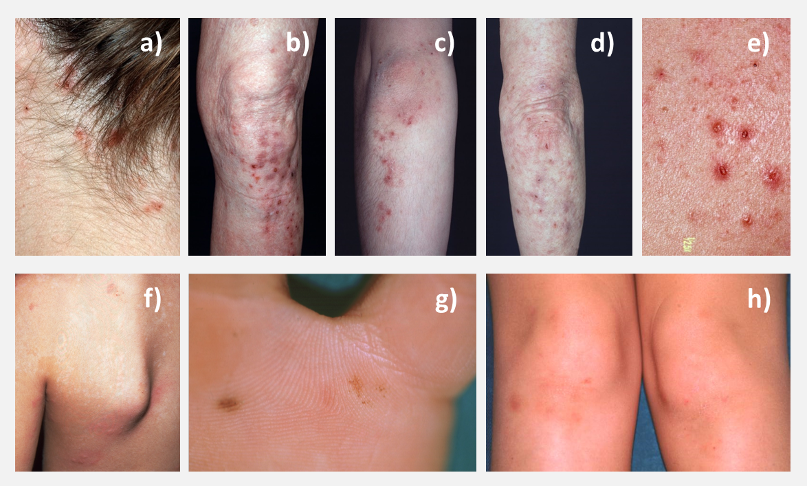
Figure 9 a to h: Several examples for clinical presentations of DHD;
a) scalp, b) knee, c) and d) elbow, e) close up-view, f) granular IgA deposits g) palmar lesions h) mild presentation in a child
Source: with courtesy from PD Dr. Christian Rose, Lübeck, Germany (a – e) and Dr. Ilma Korponay-Szabó, Budapest, Hungary (f-h)
The disease is characterized by granular IgA deposits in the papillae dermis of the skin. These IgA deposits lead to activation of the complement system resulting in an inflammatory reaction with itching small blisters. The autoantigen of DH is epidermal transglutaminase which is found in the skin at the same site as the granular IgA deposits (Sardy et al 2002; Donaldson et al 2007, Görög et al, 2021).
DH is a rare disease and only a very small group of patients with CeD will develop this skin manifestation. It usually occurs in adult patients and is uncommon in children. Males are affected more often than females (Reunala et al, 2015; Collin et al. 2017, Görög et al, 2021). Although only 10% of patients with DH report mild gastrointestinal symptoms of CeD, the majority have, at least, subclinical signs of CeD and villous atrophy (Rose et al. 2009).
The gold standard for diagnosis of DH is the detection of granular IgA deposits in the skin of patients. As all patients with DH have CeD, treatment consists of a strict and life-long gluten-free diet. The diet alone is effective in most childhood DH cases. Medical treatment with dapsone is very effective to treat the severely pruritic skin eruptions but has no effect on CeD (Reunala et al, 2015, Görög et al, 2021). However, these drugs have side effects and their use often leads to neglecting the gluten-free diet. Therefore, their prescription should be kept at the minimum.
Refractory CeD (Refractory Sprue)
If symptoms and signs of CeD do not resolve or re-occur on a gluten-free diet, the reasons are in most cases unintended gluten-intake, other gastrointestinal disorders or prolonged healing of the small bowel mucosa, which may take up to 2 years. However, there is also a severe but rare complication called refractory celiac disease (RCeD), also called refractory sprue (Verkarre et al 2003; Rubio-Tapia et al 2010). While RCeD has not been reported in children and adolescents with CeD, probably around 1% of adult patients are affected, most of them are beyond 50 years of age (Roshan et al, 2011; Rowinski & Christensen 2016; van Wanrooij et al, 2017).
The following symptoms in a patient with CeD keeping a strict gluten-free diet warrant extensive diagnostic work up: chronic diarrhoea, unintended weight loss, fever, fatigue and night sweats of unclear reasons. Based on the number and characteristics of certain immune cells (intraepithelial lymphocytes, IEL) in the mucosa, RCeD is subdivided in:
-
type I (RCD1): Marsh 3 lesions with normal types of intraepithelial lymphocytes
-
type II (RCD2): Marsh 3 lesions with an increased number (>20%) of aberrant intraepithelial lymphocytes (CD3 and CD8 negative) with pre-malignant characteristics and T-cell receptor clonality.
Patients with ReCD should be diagnosed, treated and monitored in specialist units (Felber et al, 2014). If the diagnosis is confirmed, medical therapy (in addition to the gluten-free diet) and close monitoring is needed due to the increased risk for malignancy in the small intestine (enteropathy associated T-cell lymphoma, EATL) in patients with type II RCeD.